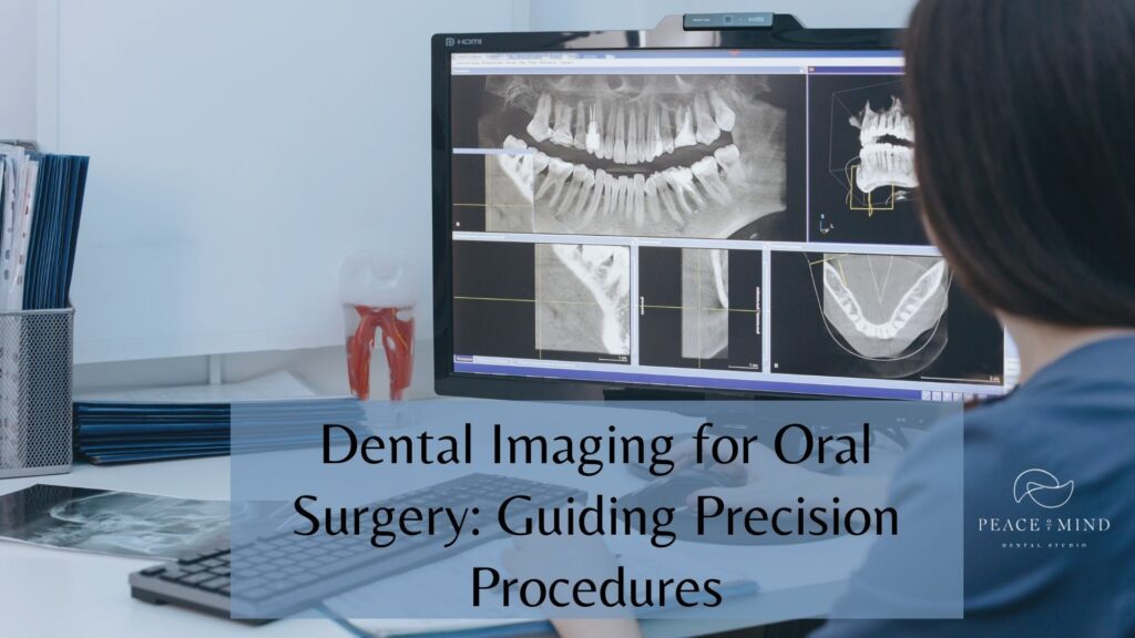
Dental imaging is the visual representation of a patient’s oral health using different diagnostics techniques and technologies. It includes detailed pictures of teeth, gums, and surrounding tissues. Dental imaging includes X-rays, CBCT scans, intraoral cameras, MRI scans, etc. These different techniques offer different advantages and detect various abnormalities of the mouth.
If you are going to the dentist for the first time and are curious about dental imaging, this blog is for you.
Importance of Dental Imaging in Oral Surgery
Panoramic radiographs, dental CT scans, periapical radiographs, etc., are different names of dental imaging, but what is it? What is its importance in oral surgery? Here are several key aspects highlighting the importance of dental imaging in oral surgery:
Diagnosis and Treatment
Dental imaging offers a comprehensive view of oral structures. Dentists can identify multiple dental issues and anomalies in a patient’s mouth, which might not be possible without dental imaging. Dental images play a crucial role in forming the right dental treatment plan.
Surgical Precision
Dental imaging is essential for identifying nerves and roots, especially during dental procedures like tooth extractions and dental implants. This technology helps minimize the risk of harming surrounding tissues and prevents potential complications.
Risk Assessment
Complex surgeries and implants can be risky. To perform successful procedures, dentists must be well-informed about dental infections, inflammations, and potential challenges. Dental imaging helps in identifying the challenges that might arise during procedures.
Informed Consent
Dental images are beneficial in communicating problems to the patients and help them understand the complexity. Patients can better understand the surgical plan and potential outcomes, allowing them to make well-informed decisions about their treatment.
Types of Dental Imaging for Oral Surgery
Dental imaging has become an indispensable tool in the arsenal of dentists. Today, various types of dental imaging techniques are employed to aid in the diagnosis, treatment planning, and execution of oral surgical procedures. Here are some of the key types of dental imaging used in oral surgery:
Panoramic Radiography
A panoramic radiograph offers a comprehensive view of the oral and maxillofacial region. It is a great tool to identify impacted teeth and bone structures.
CBCT Scans
Cone Beam Computed Tomography is the full form of CBCT scan. It is a 3D technique that offers detailed images of the oral cavity and lower face. It provides information about the bone density, volume, and anatomical structures.
Periapical Radiography
A periapical radiograph depicts one tooth and its surrounding tissues in detail. It is an X-ray type that comes in handy when your dentist needs to assess the infections of one tooth in detail.
MRI
Magnetic Response Imaging is not often used in oral surgery but is often employed to assess soft tissues, nerve locations, etc.
Intraoral Radiography
Intraoral X-rays involve small film or digital sensors placed inside the patient’s mouth. These images are helpful for detailed views of individual teeth and the surrounding bone.
How Dental Imaging Guides the Placement of Dental Implants?
A dental implant is a complex dental procedure that involves installing an artificial titanium root. Thus, dental imaging is pivotal in guiding dentists to ensure successful implants.
Here is how dental imaging contributes to the placement of dental implants:
Diagnosis and Assessment
Dental imaging helps in assessing a patient’s oral anatomy. The dentists identify bone density, quality, and potential issues before suggesting the dental implant procedure.
Treatment Planning
With the 3D view of the jawbone and facial structure, dentists can plan the optimal position of the implant location, angle, and depth and ensure that it aligns with the patient’s aesthetic requirements.
Selecting the Right Implant Size
Precise measurements from dental imaging help choose the correct implant size, ensuring that it fits securely in the available bone without damaging neighboring structures.
Monitoring Healing
A dental implant is a lengthy procedure wherein a titanium root is fixed in the jaw, and the rest of the process is performed only when the jawbone accepts the foreign root. It must integrate successfully with the bone. After implant placement, dental imaging helps in monitoring the healing process.
Dental Imaging to Get Rid of Teeth Cavity
Dental imaging helps diagnose teeth cavities. Cavities known as dental carries are small gaps in teeth by bacteria that lead to pain and sensitivity if not treated on time. Teeth cavity images help dentists notice even the most minor cavities, thus encouraging a proper cavity treatment plan.
Post-Operative Evaluation
Dental imaging is valuable for tracking the healing process and evaluating the success of surgical interventions. It provides a baseline for post-operative assessments and can reveal any complications or issues requiring additional treatment.
Final Words
Dental imaging is indispensable in oral surgery as it facilitates oral surgery, treatment planning, post-operative evaluation, monitoring healing, etc.
At Peace of Mind Dental Studio, Chandler, AZ, we use dental imaging to ensure you receive the best treatment for dental ailments.
For any dental issue, visit Peace of Mind Dental Studio in Chandler, AZ.
Frequently Asked Questions
Que: Are dental X-rays safe?
Ans: Yes, Dental X-rays are safe if radiation exposure is minimal. Dentists take precautions to ensure you are not exposed to radiation more than required.
Que: Can I have dental X-rays taken during pregnancy?
Ans: Avoid dental x-rays during pregnancy, especially during the first trimester. If it is unavoidable, communicate with your doctor about your condition so they take precautions.
Que: Are there alternatives to traditional dental X-rays?
Ans: Yes, there are alternatives, such as digital X-rays and CBCT scans, which offer detailed images and less radiation exposure.


